The laboratories have been installed in 2014 in building B5a (Physics Department). Short descriptions of the main equipment available are given below.
Helium closed-cycle cryostat and polarization microscope
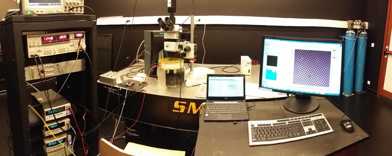
The cryostat is a custom Cryostation model designed by Montana Instruments. It can achieve temperatures between 3.6 K and 350 K with the round castle mount and the low working distance option installed. This option gives optical access to the sample through the top window of the cryostat. The temperature stability is 10 mK.
The cryostat has 20 electrical connections to the sample chamber a transport measurements can be performed with the help of K2182 nanovoltmeter, K2440 and K6221 ac/dc current sources and SR830 lock-in amplifier. A coil can be used to apply a magnetic field up to 12.5 mT at the sample position.
With the help of a garnet positioned on top of the sample, magneto-optical imaging experiments can be performed by using an Olympus BXFM modular microscope. Polarized light in sent on the sample, reflected by the garnet mirror and passes through an analyser with angular resolution of 0.1°. The polarization angle is rotated proportionally to the local magnetic field, so images of the magnetic field can be obtained, with a spatial resolution around 5 µm. 5x to 100x magnification and bright/dark field configurations are also available.
The whole setup is mounted on an actively damped optical table designed by Newport.
Wedge manual Wire Bonder
The wirebonder is an iBond5000 Series produced by Kulicke & Soffa. It is a mechanical bonder with an advanced graphical user interface. It can achieve wedge bonding on a variety of materials (Au, Cu, Pb, Al,…) with different types of wires (Au, Al, Cu, ribbon). It has a Deep Access capability, Advanced Wedge Automatic Wire Re-Feed, Semi-automatic/manual Z-mode and includes a Nikon SMZ660 Microscope 0.8-5x zoom, 20x eyepiece. It has also a built in temperature controller (for gold bonding).
Nanolithography
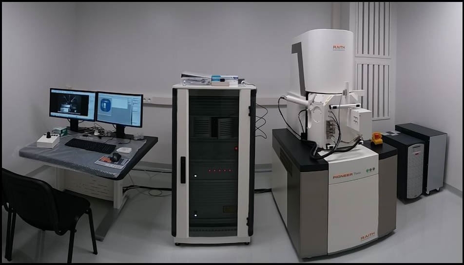
Our laboratory is equipped with an ultra high resolution electron beam lithography and scanning electron microscope imaging system RAITH Pioneer Two.
A highly precise laser interferometer controlled stage enables state-of-the-art results in exposures, which require excellent pattern placement (2 nm), stitching (< 60 nm) and overlay performance (< 50 nm). This system allows to easily write samples down to approximately 10 nm size. The energy of the electron beam can be tuned from 100 V to 30 kV (with 10 V steps). Different apertures sizes can be chosen, from 7 µm to 120 µm, allowing to have a beam current from 5 pA to 20 nA. The system allows for full coverage of 2'' wafers, i.e. 50 mm by 50 mm travel range. The system is equipped with an Everhart-Thornley Secondary Electron detector, an In-Lens detector allowing high resolution electron imagery below 20 kV, and a backscattered electron detector for Z-contrast. Integrated platforms allows sample rotation and tilt for observation as well as in-situ combined electrical measurements and inspection of nanodevices. The EBL system is located in an over pressured room with controlled temperature stability (± 0.1 °C) and humidity (< 60 %), and with reduced particle pollution through high performance HEPA filters.
Physical vapor deposition
Multipurpose system from Angstrom Engineering for thin film synthesis by physical vapor deposition (PVD). This tool consists of a vacuum chamber equipped with targets for electron-beam evaporation and radio-frequency sputtering. Temperature of compatible substrates, either wafers up to 6″ diameter or small square pieces, can be controlled from room temperature to 600°C. Deposited thickness homogeneity is enabled by the rotation of the substrate stage. Deposition rate is monitored by quartz crystals and ambient gas atmospheres can be set by mass-flow controllers. The whole system is managed by a dedicated PLC-coupled software and is integrated into an inert-gas glovebox assembly. The range of materials which can be deposited with this PVD equipment includes : Al, Cu, Nb, Ni, Au, SiO2, Al2O3, NbTi, ZnO, etc.
Sample preparation facilities
A chemical room for sample preparation equipped with a ducted high performance Acela Frontier fume hood, a laboratory refrigerator Labo 100 from Kirsch (2°C-20°C), programmable universal oven Memmert UF75 (300°C), ultrasonic bath, high precision industrial hot plate Unitemp HP-155 with vacuum fixing (350°C), spin coater Laurell, and a compact rapid thermal annealing system.
Atomic Force Microscopy
The laboratory is furnished with two Atomic Force Microscopes. The multimode-8 AFM enables users to conduct topography scanning in both tapping and contact modes, as well as perform Magnetic Force Microscopy (MFM) measurements. The scanning size is restricted to 10×10 µm², with a z-sensing range of 5 µm.
The Drive AFM offers additional modes, including EFM, c-AFM, EC-AFM, KPFM, and force spectroscopy. It comes with a lateral scan range of 100×100 µm² and a vertical range of 20 µm. Moreover, the microscope is equipped with a magnetic field generator capable of producing a maximum field strength of 800 mT.
Magneto-dynamics platform
The magneto-dynamics platform is capable of conducting a wide range of experiments, including broadband FerroMagnetic Resonance (FMR), spin-torque FMR (ST-FMR), and Propagation Spin Wave Spectroscopy (PSWS). This setup consists of a Vector Network Analyzer (VNA) with a frequency range spanning from 5 kHz to 20 GHz, a magnetic field generator capable of producing a maximum field strength of 500 mT, and two picoprobes.